
Microsurgery Explained: Procedures, Instruments & Applications in 2025
In today's surgical miracles, cancer surgeons replace gaps left after tumor removals with flaps, plastic surgeons replace burnt skin with full thickness grafts, trauma surgeons replant severed fingers or limbs!
All the above are possible with the crucial support of micro surgery.
Microsurgery is one of the most elegant and precise domains in the vast landscape of surgery.
Microsurgery procedures are often invisible to the naked eye and are conducted under a high-powered microscope. This important adjunct to surgery plays a pivotal role in many complex operations.
Microsurgery is not a standalone surgical specialty. Rather, it is a powerful technique used alongside a wide range of surgical procedures, some of which are mentioned and above. And, the success or failure of every surgery where microsurgery is used, depends on the success of the microsurgery procedure.
This article explains:
- What microsurgery is
- How it works
- What instruments it uses
- The kinds of procedures it enables
-
Why it's vital for modern surgical outcomes
What is Microsurgery?
Microsurgery means very minute surgical procedures performed on very small structures, such as blood vessels, nerves, and lymphatics, and which must be done under magnification, under a surgical operating microscope. The goal of microsurgery is to enable precise manipulation of these tiny components and to repair or reconstruct tissues.
One aspect of microsurgery is microvascular surgery, in which small blood vessels are joined to establish blood flow. We will learn more about it later.
Microvascular surgery constitutes 78% of all microsurgeries.
A Brief History of Microsurgery
The practice of microsurgery began in the 1960s with pioneering efforts in reattaching severed limbs and reestablishing circulation.
A finger or a limb which is cut off, can be joined to its stump by joining the bones, muscles, and skin with plates, surgical staples, and stitches. But it is not enough.
This joining will not succeed, unless and until, blood supply to the newly joined part is established from the local blood vessels!
This is what microvascular surgery does.
It joins the blood vessels of the newly attached part to local blood vessels.
Since the 60's, micro surgery and micro vascular surgery have expanded into multiple fields such as:
- Cancer surgery
- Plastic plus reconstructive surgery
- Neurosurgery
- Ophthalmology
- Otolaryngology
-
Urology
Core Principle of Microsurgery: Anastomosing Small Blood Vessels
The major work done in microsurgery is vascular anastomosis, which means the precise joining of blood vessels with an aim of restoring blood supply to the newly joined part.
This is essential in organ transplantation, free flap reconstructions, and limb or finger replantations. Vessels as small as 1 mm in diameter can be anastomosed with stunning precision.
A successful anostomosis ensures healthy blood supply to the newly joined part. This in turn helps tissues to survive in places where their native blood supply has been lost.
78% of microsurgeries are done to anastomose small arteries or veins.
How Anastomosis Works
Anastomosis means a surgical connection between two structures, usually blood vessels or tubular organs, to allow flow between them.
Apart from blood vessels, anastomosis is also done in much larger organs like Intestines, Ureters, Bile ducts, Fallopian tubes, Oesophagus, Trachea, Bronchi etc.
In all of the above, anastomosis ensures continuity of flow whether it’s blood, air, urine, bile, or intestinal contents.
Being large organs, it is somewhat easy to anastomose them as compared to small blood vessels barely 1 mm in diameter! Most of the above don’t need microsurgery.
But, how is it possible to stitch such small vessels together, and still have them function without leaking? How to ensure blood flow occurs through the skillfully made anastomosis?
How is Anastomosis done?
The precision in Anastomosis is possible because of two things:
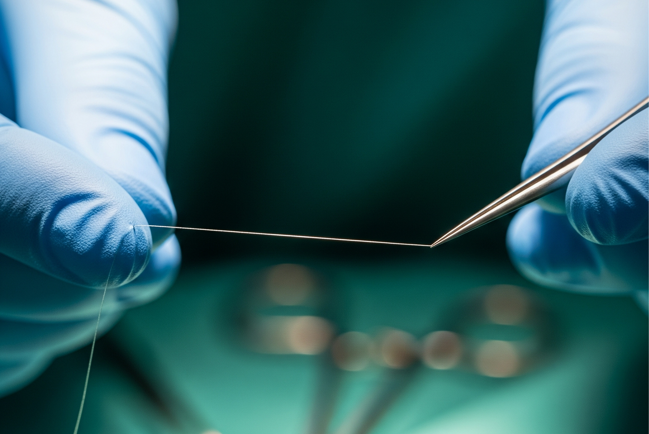
- Accurate end-to-end approximation: Surgeons align the intimal (innermost) layers of both vessels exactly.
- Microsurgical suture technique: Sutures are placed using ultrafine threads, typically 9-0 to 11-0 nylon or prolene. The suture bites are tiny and spaced evenly to avoid gaps.
- Watertight seal: The blood pressure itself helps seal the vessel from the inside if alignment is perfect. A good anastomosis creates a watertight seal, and the vessel heals quickly due to its rich endothelial lining.

Critical Surgeries That Depend on Microvascular Anastomosis
There are certain surgeries where success entirely depends on restoring blood supply to the tissue being implanted, grafted, or reattached. These surgeries are only possible because of microsurgical techniques, specifically vascular anastomosis (the precise joining of tiny blood vessels)
- Flap surgery: means moving living tissue with its own blood supply from one part of the body to another to repair or reconstruct a damaged area.
- Reconstructive surgeries: such as breast reconstruction after a surgical removal of a breast, mostly because of a tumour.
- Skin grafting: especially full-thickness grafts in cases of severe burns.
- Reimplantation of limbs or digits: to restore function and viability.
- Tumor resections with reconstruction: where large areas require vascularized tissue to fill the gaps left by tumor removal.
- Free tissue transfers: including the bone fibula with skin and muscle.
In essence, tissue viability and healing in nearly all surgical specialties depends heavily on a healthy, well-maintained blood supply.
Other Surgeries that require Microsurgical Techniques
Microsurgery is also performed in procedures where vascular anastomosis is not the primary goal. These are the remaining 22% of all microsurgeries.
Examples are:
- Nerve repair, especially in cases of brachial plexus injuries
- Nerve injuries of the fingers, microsurgical techniques are used to align and suture tiny nerve fibers.
- Reconstructive surgery after cancer, trauma, or congenital defects often involves meticulous dissection and tissue handling under a microscope.
- Lymphedema surgery, particularly lymphaticovenular anastomosis (LVA), targets lymphatic vessels rather than blood vessels.
- Eye surgeries such as retinal or corneal procedures also use microsurgical tools.
-
Ear surgeries, including cochlear implants, rely on microsurgery to handle minute, delicate structures.
List of Surgical Domains that involves Microsurgeries
This is a list of surgical domains where microsurgery is a vital part of the main procedure:
1. Neurosurgery:
- Microvascular decompression
- Tumor removal near cranial nerves
- Brachial plexus repair
2. Ophthalmology:
- Retinal surgery
- Corneal transplantation
- Glaucoma surgeries
- Cataract extraction
3. ENT (Otolaryngology):
- Microsurgery for vocal cord polyps
- Cochlear implantation
- Ossiculoplasty (middle ear surgery)
4. Urology and Gynecology:
- Vasectomy reversal
- Tuboplasty
- Varicocele repair
5. Orthopedic and Hand Surgery:
- Tendon and nerve repair
- Limb salvage procedures
- Carpal tunnel release under micro-visualization
Instruments Used in Microsurgery
A regular pair of scissors or forceps will hardly be useful in microsurgery. As can be guessed, microsurgery depends on very small specialized tools, the most important of which is, obviously, the operating microscope.
However, every instrument is important, and is designed for precision, minimal trauma, and the control of each instrument is optimised for use under an operating microscope.
Microsurgical instruments are:
- Operating Microscope: A high-powered binocular microscope providing magnification ranging from 5 times to 40 times.
- Microsurgical Needles and Sutures: Needles smaller than a grain of rice; sutures are 9-0 to 11-0 in size.
- Microscissors: Ultra-fine scissors for sharp dissection.
- Microforceps: Delicate forceps for handling nerves and vessels.
- Microvascular Clamps: Temporary clamps to block blood flow during anastomosis.
- Needle Holders: Extremely precise holders suited to the tiny needles used.
- Vessel Dilators: Used to gently open and prepare vessels.
- Approximators: Devices to hold vessels in place during stitching.
- Laser Tools: In some eye and nerve surgeries.
- Electrocautery Tools with Fine Tips: For delicate coagulation.
- Tissue Stabilizers: Help reduce tremors during intricate moves.
- Microsurgical Loupe Glasses: Portable magnification (for procedures not requiring the operating microscope.
It is needless to state that each tool must be handled with exceptional dexterity. Even the slightest hand tremor in a surgeon’s hand can result in an adverse result.
How We Help at Shira
At Shira, we design microsurgical instruments, like our innovative 3-jaw clamp, to meet the demands of vascular precision and surgeon comfort.
Limits and Possibilities: How Small is Small?
The best microsurgeons in the world have performed anastomoses on vessels as small as 0.3 mm. Some surgeons can repair nerve fibers 10 microns in diameter. That’s one-tenth the width of a human hair. With robotic assistance, this threshold will continue to fall.
Human tissue is not rigid, and the surgical field constantly shifts with pulse and respiration. A microsurgeon must compensate for all these factors while working in magnified space. The success relies not only on visual magnification, but also on the surgeon’s motor skill, tactile feedback, and detailed anatomical understanding.
The Training Behind the Art
Microsurgeons undergo rigorous training beyond general surgical skills. This includes:
- Hours of practice on synthetic models and animal vessels
- Mastery of hand-eye coordination
- Understanding of tissue mechanics at micro levels
- Humility and patience—errors at this scale are unforgiving
Many microsurgical training centers use simulation labs for skill development.
Some even require the candidate to perform perfect anastomoses under a microscope in a live animal before they are allowed to operate on humans.
Microsurgery and Robotics
Robotic systems like the da Vinci Surgical System have taken microsurgery to the next level. These systems provide ultra-fine movement translation, eliminate tremor, and allow for remote surgery.
However, human dexterity and judgment are still irreplaceable. The robot can aid but not replace the artistic judgment of a skilled surgeon’s hands.
Conclusion: The Vital Hero of Modern Surgery
People talk about cancer surgeons who successfully operated on a mouth cancer, they praise plastic surgeons for thick skin graft on major burns or for reattaching a limb. But no one mentions microsurgeons, even though they have done the anastomoses and helped establish a viable blood supply to the newly attached flap or skin!
Yet, microsurgery is one of the most critical foundations in modern surgical success. Without it, limb reattachments, free tissue transfers after cancer surgeries, and delicate nerve repairs would be impossible.
It is not a discipline in isolation but a technique, a set of skills, and a world of instruments used across specialties. Whether giving a new face to an accident victim or restoring sight to a blind eye or reconstructing a jaw removed due to cancer, microsurgery is the quiet hero that makes it happen!
The future of microsurgery lies in hybrid techniques—merging the surgeon’s hand with robotic precision and AI-guided planning. But its soul will always be the human hand, trained to move with the grace and certainty of a maestro.