
Joining 1 mm Blood Vessels: Surgeries That Chooses Anastomosis
Believe it or not, small blood vessels, both arteries and veins, which are as narrow as 1 mm [or even narrower!] are joined end to end as a vital part of many surgeries. In these delicate procedures, handsewn methods remain a traditional approach valued for their accuracy and control in microsurgery. Additionally, full thickness sutures are sometimes used to ensure the integrity of the vessel join, especially in challenging cases.
List of Surgeries That Depend on Tiny Vessel Joins
| S. No. | Types of Surgeries | Brief Explanation |
|
1 |
Digital replantation |
Surgical reattachment of fingers or toes that have been completely amputated. |
|
2 |
Fibula free flap reconstruction |
Transfer of the fibula bone with blood vessels to reconstruct long bone defects, often in limbs. |
|
3 |
Free flap breast reconstruction |
Using tissue from other parts of the body (like abdomen or thigh) with its blood supply to reconstruct the breast. |
|
4 |
Ear replantation |
Microsurgical reattachment of a completely amputated ear, preserving function and appearance. |
|
5 |
Nasal reconstruction |
Rebuilding of the nose using grafts or flaps to restore shape and breathing function after trauma or cancer. |
|
6 |
Vascularized nerve grafts |
Transplanting nerve tissue with its own blood supply to bridge nerve gaps and restore function. |
|
7 |
Free fibula jaw reconstruction |
Using the fibula bone and its vessels to reconstruct segments of the jaw, often after tumor removal, Commando. |
|
8 |
Tongue reconstruction |
Reconstructing tongue defects using tissue flaps to preserve speech and swallowing functions. |
This list is only indicative. The real list is long, many more surgeries are not included in this list for the sake of brevity. Thus, microsurgical anastomosis, which means joining small blood vessels end to end, is needed quite often.
Let’s follow a team of surgeons doing a real life Commando Surgery to understand microsurgery.
A Day inside the OR: Understanding Commando Surgery
Commando Surgery removes advanced mouth cancers in the jaw, tongue, or throat. After excision, the remaining ends of healthy tissue are carefully prepared for reconnection. Surgeons reconstruct the area using transplanted tissue (like the leg bone called fibula, with muscle and skin), reconnecting tiny blood vessels under a microscope to restore blood supply [aka anastomosis]. Achieving a successful anastomosis is critical to ensure the viability and optimal healing of the reconstructed tissue. It’s a major, life-saving procedure for aggressive tumors. This long, complex surgery lasts for 10–14 hours. It restores eating, speaking, and facial appearance.
When the Clock Starts at 6 AM: Anatomy of a High-Stakes Surgery
Unless the surgical team leader is a beast, complex surgeries don’t succeed. Most surgeons are sticklers to punctuality for long lasting planned surgeries like commando surgeries. If they say 6 am, they often make the first incision at that very moment.
Needless to say, before the first incision, many things have happened.
-
The operating room [OR] staff and the anaesthetist come one hour before the cancer surgeon and his associates.
-
They have checked that the patient has fasted overnight, the surgical areas have been shaved, his regular meds have been given, and his vitals are within normal limits.
-
Then the OR team and the anaesthetist do their work, which includes giving anaesthesia and draping the part.
-
The surgical trolley with about 700+ instruments was made ready and the special air conditioning of the OR was activated one hour ago.
During these meticulous preparations, specialized instruments are arranged for the reconstructive steps, and surgeons may also use their index finger to gently palpate and confirm the alignment or patency of joined tissues during the procedure.
Commando surgeries need cancer surgeons, plastic surgeons and microsurgeons, apart from the OR staff, anaesthetists, and critical care specialists for post operative care if needed. Often the latter two are the same person.
Today’s case is formidable:
A commando surgery, where half of the patient’s jaw and tongue are to be removed due to an aggressive carcinoma. A fibula free flap, will be harvested from the leg, and a portion of the pectoralis major muscle from the chest will be meticulously shaped and positioned to reconstruct what was lost through surgery.
But even after all the structural artistry, one element remains critical, restoring the blood supply to the newly attached body parts.
When the clock in the operating room ticks past the eleventh hour:
-
Cancerous jaw and tongue removed
-
Fibula with muscle and skin harvested
-
Devastated facial area ready for reconstruction
-
Microsurgical flap to be attached and vascularized
This is a crucial part of the surgery. Remember, a part of the muscle, skin, and bone from the leg and chest has been removed, and is about to be attached to the jaw.
This attachment doesn’t mean just sewing them together or attaching the bones with screws and plates. It also means anastomosing the local blood vessels to the native blood vessels of the newly attached part, which is known as the ‘flap’.
The newly attached flap needs a blood supply, and that is what end-to-end anastomosis achieves, by joining the arteries to bring in the fresh blood supply to the flap and the veins to drain away the deoxygenated blood from the flap.
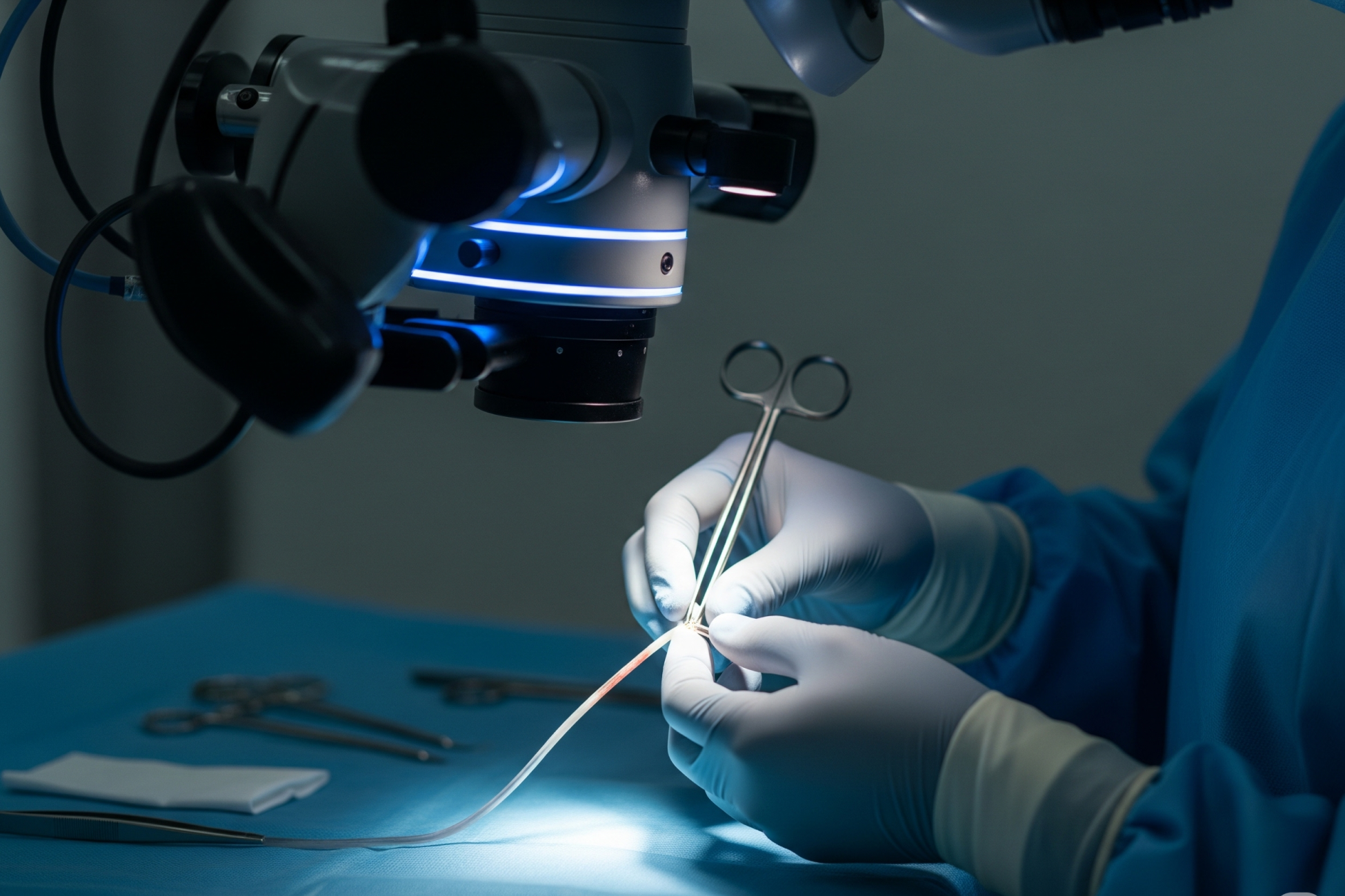
A sterile silence lingers in the OR, broken only by the hum of the laminar air flow of the OR, subtle clinking of titanium and stainless steel instruments, and the rhythmic beeping of the anesthesia monitor. The microsurgeon, working through an operating microscope, is clad in a gown and micro-gloves. He is completely immersed in his task, joining blood vessels so that the newly attached parts will be vascularised and survive, and thrive.
Brief explanation of Micro-gloves?
Yes, Micro-gloves, which are special gloves used in microsurgery. They differ from regular surgical gloves in key ways to facilitate the extreme precision and delicacy required when operating on tiny blood vessels, nerves, or tissues.
Till now, this surgery was done under the unaided eyes [eyeglasses not counted] of cancer and plastic surgeons. For joining 1 mm wide blood vessels, the microsurgeon uses the operating microscope. It gives a magnification of five times to forty times, called 5x to 40x.
Microsurgical Vascular Anastomosis: A Battle Against Time and Tension
In reconstructive microsurgery, the success of a free flap hinges on connecting tiny arteries and veins, some just 1 millimeter or less in diameter, with immaculate precision. These are typically sutured end-to-end to re-establish blood flow from the body to the transplanted tissue, though vessels can be sewn or stapled depending on the specific requirements of the procedure. This process is known as vascular anastomosis.
To perform this task, the vessels are temporarily clamped, using small, delicate instruments typically just 2 to three centimeters long. These vascular clamps hold the two ends of the two blood vessels and halt the blood flow, allowing the surgeon to suture in a relatively bloodless field. In recent years, the stapled technique has also emerged as an alternative to traditional suturing in certain microsurgical contexts.
But here lies the challenge: how to hold these minuscule, flexible vessels in the correct position—everted, open, and stable, so that the suture needle pierces only the vessel wall once, and does not inadvertently go through both walls (known as “back-walling”)? The consequences of a poor anastomosis are immediate and catastrophic: thrombosis, flap failure, tissue necrosis. In short, failed surgery!
Vessel Eversion: A Crucial Step
Eversion is like the first step of rolling up the sleeve of your full-sleeved dress shirt. The inner side of your shirt sleeve comes out.
Eversion of the tiny blood vessel edges is necessary when joining them. Their inner sides have to be curled out a bit so that their inner layers touch each other, and then they can be sewn or sutured together for a firm join.
Eversion brings a small part of the delicate intimal layer (the innermost lining of the blood vessel) to the outer side. When both vessel ends are everted and successfully sutured inner layer to inner layer, the flow of blood is smooth. With proper eversion, the possibility of the edge of the blood vessel curling inwards is prevented. This smooth join reduces turbulence and has less risk of forming a blood clot within it.
It’s like ensuring a road junction is smooth and aligned—no bumps, no sharp turns, no obstructions, at the junction. Similarly, in intestinal anastomosis, precise alignment and secure suturing of the bowel ends is essential to ensure a viable, tension-free join and successful healing.
Without proper eversion, the risk of sutures inadvertently piercing both the front walls and the back walls of the vessel increases dramatically. Such errors can collapse the lumen, obstruct flow, or form clots, all of which doom the surgery.
Eversion isn’t just a desirable feature—it’s a non-negotiable ‘must’!

Three-Jaw Clamp Mechanics, Explained
This brings us to a quiet revolution in microsurgical technique: the advent of a three-jaw clamp.
Unlike traditional two-jaw clamps that only hold the vessel in one axis, the three-jaw design introduces a game-changing dimension:
-
a gently tensioned three-point stability
-
consistent eversion
-
enabling reliable of the blood vessels being joined, or anastomosed.
Why Two-Jaw Clamps Weren’t Enough
One clamp of the three-jaw structure clamps the vessel, and the second clamp of the three-jaw structure holds the everted edge of the blood vessel!
So, of the three jaws of the three-jaw clamp, the middle and lower jaw hold the blood vessel, the middle jaw acts as a fulcrum around which the surgeon everts the inner side of the blood vessel. The top jaw holds the everted edge of the blood vessel gently, yet firmly, enabling the microsurgeon to suture it with the similarly clamped and everted edge of the other blood vessel.
So now the clamps on either blood vessel are holding the tiny blood vessels while their edges are everted.
This is a position that is ideal for suturing the two blood vessels from the back side to the front side. This is a mini revolution.
Because, before the three-jaw clamp was invented, microsurgeons used two-jaw clamps which offered no eversion, managed to evert the edge of the vessels with their forceps, scissors, and needles, and sutured the blood vessels, beginning from the front side.
Then they flipped the two clamps holding the two blood vessels to sew the back side of the vessels. This was a very risky step as flipping the delicate vessels could injure them.
Three jaws:
-
Middle & lower jaw → hold vessel
-
Middle jaw = fulcrum for eversion
-
Top jaw → gently holds everted edge
Old Method: Flip clamp to sew back side → Risky!
New Method: No flipping, no injury, precise suture
The Impact on Surgical Flow and Outcomes
In microsurgery, where even the patient’s breathing movement or a slight tremor in the microsugeon’s hand can adversely affect outcomes, flipping the clamps 180° does seem somewhat violent and savage, in a surgery so delicate!
The three-jaw clamp, on the other hand, ensures that the blood vessel stays properly everted throughout the procedure. This minimizes, almost obviates, the dreaded risk of back-walling, or accidentally suturing both walls of the blood vessel, and ensures clean, smooth flow through the new join, when the clamps are finally removed.
Echoes of a Post-Operative Conversation in the OR
In the quiet moments after the clamps come off, the fibula flap has ‘taken’. The blood vessels are pulsating. The flap muscle is pink. The pulse oximeter on the graft gives a perfect reading. The blood flow has resumed. As part of the immediate post-operative evaluation, the surgical team also carefully monitors for any signs of anastomotic leak, a critical complication that requires early diagnosis and management. The job was done right.
The surgeon finally leans back, his smile is hidden within his surgical mask, but his eyes sparkle. Sweat lines the edge of his brow under the surgical cap. He nods to the anesthetist, who’s been monitoring vitals for hours.
“Happy?” she asks with a smile.
He replies, finally allowing himself a moment of levity, “Couldn’t be better. There was a time I used two separate clamps, and it felt like holding jelly with chopsticks.”
She chuckles.
“This new clamp, you know the one,” he says, gesturing toward the surgical tray. “Changed everything. That third jaw makes all the difference. Keep the lumen open. Let me see what I’m sewing. I don’t worry about catching the back wall anymore.”
There’s a moment of appreciative silence between them. It’s not just about instruments, it’s about trust, lives, and the quiet victories of engineering working hand-in-glove with human hands.
“Don’t relax, anyone,” the beast surgeon shouts. “Still a long way to go. But I will have a cup of coffee before I suture the muscle and skin. Remember, black, no sugar!”
Sometimes, the greatest advances in medicine aren’t loud. They’re quiet, precise, and barely a few centimeters long.
Just like the three-jaw clamp.
Science, Precision, and the Role of Engineering
What often goes unspoken in surgical innovation is the quiet role of engineering. Medical tools are not just passive accessories—they are co-surgeons. The three-jaw clamp reflects this beautifully. Its design was not born in a vacuum but out of the real, daily struggles of microsurgeons trying to suture 1-mm arteries under a microscope.
Innovations like the three-jaw clamp have significantly improved outcomes in a wide range of surgical anastomoses, where precise connection of vessels or other structures is critical for successful medical interventions.
This clamp’s success lies in understanding the biomechanics of the vessel wall, the physics of tension and traction, and the physiology of blood flow. It allows the intima to meet the intima, lumen to stay patent, and blood to flow uninterrupted, just as nature intended.
Flow Patency – The Final Proof of Success
All efforts in microsurgical anastomosis lead to a single moment, the release of the clamp, and beginning of blood flow through the new anastomosis. That’s when the surgeon holds his breath. The vessel should expand, not collapse. The blood should rush through, not clot. The newly grafted tissue should become pink, not pale. Flow patency is the final and ultimate proof of success.
The three-jaw clamp, by facilitating proper eversion and precision suturing, dramatically improves the chances of this happy ending.
Patient Outcomes: Measuring Success Beyond the OR
The true measure of success in anastomosis surgeries—be it intestinal anastomosis, colorectal anastomosis surgery, or vascular procedures—extends far beyond the final suture in the operating room. Patient outcomes are the ultimate benchmark, and one of the most critical indicators is the rate of anastomotic leakage. This complication, where the surgical connection between two structures fails to seal completely, can have serious consequences for recovery and long-term health.
Research, including prospective randomized trials, has shown that the choice between stapled anastomosis and hand-sewn techniques can influence the risk of leakage. For instance, some studies have found that stapled anastomoses may offer a lower rate of anastomotic leakage compared to traditional hand-sewn methods, especially in colorectal surgery. However, outcomes are also shaped by factors such as the patient’s overall health, the presence of conditions like diabetes mellitus, and the use of medications that affect healing or blood clotting.
At leading institutions like Virginia Mason Medical Center, a general surgery practice specializing in complex procedures such as breast cancer surgery and colorectal anastomosis surgery, a multidisciplinary approach is key. Surgeons collaborate closely with anesthesiologists, nurses, and other specialists to ensure meticulous preoperative planning, precise surgical technique, and vigilant postoperative care. This comprehensive strategy helps optimize outcomes, reduce complications, and support patients on their journey to recovery. By combining evidence-based practices with the latest advances in surgical technology, teams strive to deliver the best possible results for every patient undergoing anastomosis surgery.
Looking Ahead
As medicine becomes more interdisciplinary, we are witnessing a fusion of surgical insight and mechanical ingenuity. The three-jaw clamp is a testament to this, where a small shift in design led to a giant leap in outcomes.
-
For doctors, it's a better tool.
-
For engineers, it's a triumph of precision.
-
For patients, it's the difference between healing and surgical failure.
-
For investors and grant donors, it’s proof that even in saturated fields like surgical instrumentation, meaningful innovation is still possible.